
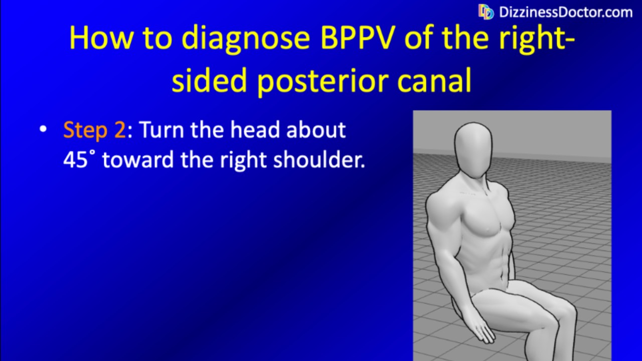
Diagnose BPPV of the posterior canal
Treating BPPV without a confirmed diagnosis is a gamble. Each canal variant produces its own characteristic pattern of eye movements — and each requires its own specific maneuver. A treatment targeted at the wrong canal won't just fail; it may make symptoms worse. In this course, we teach you to identify posterior canal BPPV with confidence: we display and explain the diagnostic eye movements that confirm the diagnosis and distinguish it from other BPPV variants, so that when treatment begins, it begins in the right direction.
This course is designed for three audiences. For patients who have already been evaluated but are still symptomatic, it offers a window into what the correct diagnostic process looks like — and whether it was followed. For clinicians who want to sharpen their nystagmus recognition skills, it provides the visual grounding that textbooks and most online resources simply don't offer. And for anyone preparing to take our treatment course, it provides the diagnostic foundation that makes treatment selection rational rather than arbitrary.
If you already know your diagnosis and are ready to move straight to treatment, our treatment-only course covers the Epley maneuver at a lower cost. But if there's any uncertainty about which canal is involved — or which side — this is the right place to start.
This material is intended for educational purposes only, and does not substitute evaluation by a qualified medical professional.
In developing this course we reviewed literature (so that you don't have to)!
- Bastani P, Phillips V, Rieiro H, Otero-Millan J, Zee D, Newman-Toker D, Tehrani A (2025) Feasibility of Using Smartphone Eye-Tracking for Self-recording Positional Tests. Digital Biomarkers: 1-11. doi: 10.1159/000545720
- Fife TD (2012) Positional dizziness. Continuum (Minneap Minn) 18: 1060-85. doi: 10.1212/01.CON.0000418376.80099.24
- Melliti A, van de Berg M, van de Berg R (2023) Capturing nystagmus during vertigo attacks using a smartphone: adherence, characteristics, pearls and pitfalls. J Neurol. doi: 10.1007/s00415-023-11965-y
- Phillips V, Bastani PB, Rieiro H, Hale DE, Otero-Millan J, Zee DS, Newman-Toker DE, Tehrani AS (2025) A Pilot Study of Smartphone Eye Tracking for Detection of Positional Nystagmus. Digit Biomark 9: 124-129. doi: 10.1159/000547008
- Shah MU, Lotterman S, Roberts D, Eisen M (2019) Smartphone telemedical emergency department consults for screening of nonacute dizziness. Laryngoscope 129: 466-469. doi: 10.1002/lary.27424
- Teixido M, Woods O, Kung B, Seyyedi M (2016) A 3D benign paroxysmal positional vertigo model for study of otolith disease. World J Otorhinolaryngol Head Neck Surg 2: 1-6. doi: https://doi.org/10.1016/j.wjorl.2016.02.002
- Traboulsi H, Teixido M (2021) BPPV Viewer: A downloadable 3D BPPV model for study of otolith disease. World Journal of Otorhinolaryngology - Head and Neck Surgery 7: 34-39. doi: https://doi.org/10.1016/j.wjorl.2018.10.001
- von Brevern M, Radtke A, Lezius F, Feldmann M, Ziese T, Lempert T, Neuhauser H (2007) Epidemiology of benign paroxysmal positional vertigo: a population based study. J Neurol Neurosurg Psychiatry 78: 710-5. doi: 10.1136/jnnp.2006.100420
